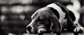
Corneal ulceration is called ulcerative keratitis – this serious eye disease that can lead to severe consequences, up to complete blindness of the pet. This pathology detected in both humans and dogs. But no matter who is patient, the main thing that the patient received timely medical help. What threatens corneal ulcer in dogs and how is this treated disease?
Content
- 1. What is ulcerative keratitis
- 2. Types of erosion and the causes of their occurrence
- 3. Symptoms of corneal ulcers in dogs
- 4. Diagnosis of the development of corneal ulcers in dogs
- 5. Treatment of corneal ulcers
What is ulcerative keratitis
The corneal layer is the front of the sheath of the organ of vision, including several layers:
- upper – epithelial, is a protective membrane of an organ vision;
- then follows the stroma – the basis of the entire cornea;
- Dessemeth membrane (desmemetova membrane) – posterior border wall;
- posterior epithelial layer – corneal endothelium, it supports mild dehydration of the eyeball.
In normal condition, the cornea has a smooth, transparent, without any roughness, the surface in its layers there are no vessels circulatory system. And since it contains a huge amount nerve roots, then this area is characterized by increased sensitivity. 
Corneal ulcerative lesion affects its uppermost epithelial layer. If we draw an analogy with skin damage, then an ulcer is a scratch, but not of the skin, but of the corneal layer, and this pathology is considered more dangerous.
The condition of a sick animal suffering from an ulcer is aggravated due to the abundance of nerve endings in the cornea, this form keratitis is accompanied by intolerable soreness. Pain knocks out a pet from a normal rut, does not allow him to eat normally, relax, causes insomnia. This condition quickly leads to a nervous and physical exhaustion of the dog.
Amid damage, degradation occurs rather quickly. epithelial layer of the eye, the body loses reliable protection against various infectious pathogens. Ulcerative keratitis is serious pathology, the complication of which is often bacterial infection of the eyes and subsequent blindness.
Types of erosion and the causes of their occurrence
There are several types of ulcerative keratitis. First of all, in depending on the cause, the disease may be infectious or non-infectious. The occurrence of infectious ulcers is often associated with viral, bacterial, fungal lesions of the cornea. Besides, damage can be caused by infection with parasites.
Such peptic ulcer disease is difficult to treat and very often recurs. Most often, an infectious ulcer causes staphylococcus, streptococcus, Pseudomonas aeruginosa, herpes virus, coronaviruses, Koch’s wand. In addition, pets Chlamydial infection can cause ulcerative keratitis.
With non-infectious keratitis, factors provoking the appearance of ulcerations on the corneal layer, the following may appear status:
- pedigree predisposition – most often eye pathology develop in dogs with bulging eyes (brachycephalus) – Pekingese, Japanese Chin, Shih Tzu, Boston Terriers, French Bulldogs, Great Danes, Labradors, etc .;
- hit of a foreign body on the cornea and third eyelid;
- a change in the position of the eyelids (inversion);
- dry eye syndrome;
- burn of the mucous membrane of the eye – chemical, thermal, ultraviolet and others;
- violation of eyelash growth (dystrichiasis, cilia) that begin scratch, rub the surface of the cornea;
- limbal corneal insufficiency;
- decrease in local immunity.
Also, experts distinguish superficial and deep ulcerative keratitis:
- superficial pathology – the lesion affects the epithelial layer and stroma of the cornea;
- deep pathology – ulcerations spread to all layers stroma. With the progression of the disease, the pathological process penetrates into the thickness of the cornea, affecting the discemet membrane, which threatens perforation of the anterior membrane of the eyeball;
- descemetocele – a condition in which a complete perforation of the corneal layers when through damage and reaches to the Descemet layer.
Special attention should be paid to chronic erosion. cornea, in which damage does not heal for a long time. Pathology is called endolent erosion or ulcer boxers. To group The risk of this pathology includes representatives of the following breeds: boxer, dachshund, spaniel, spitz, etc. Most often, the disease diagnosed in animals older than 5 years.
The peculiarity of keratitis in this case is that damage can not heal for weeks, and no visible there are no reasons for this, and the drugs used do not give positive effect.
The cause of the disease lies deep – there is a failure of cell contact epithelium with cells of the basement membrane, therefore normal recovering epithelial tissues cannot be fixed on membrane, and their desquamation occurs. Naturally, in such Under conditions, the damaged area simply has nothing to close.
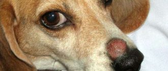
Quite often, ulcerative keratitis does not have pronounced signs, and the owner of the dog does not notice that his pet is sick. But with the disease progresses over time, the dog begins to feel severe pain and discomfort. At the first symptoms of the disease, the dog should be shown to the veterinarian.
Dog corneal ulcer symptoms
When is the dog owner concerned about the condition of the pet? IN acute stage, corneal erosion is accompanied by increased secretion lacrimal fluid and photophobia.
The pet makes constant attempts to rub his eyes with his paws, than else exacerbates the condition of the visual organ – in this situation injury to the damaged organ and secondary infection of existing wounds. The conjunctiva turns red spasms of the eyelids. 
Gradually, existing sores become more noticeable if If pathology is not treated, then a perforated ulcer may occur. A complication of this condition is panophthalmitis – acute purulent inflammatory process that affects all tissues and membranes visual organ or loss of internal parts of the eye.
Diagnosis of corneal ulcer in dogs
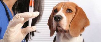
In case of eye diseases in dogs, the owner is advised to contact to a veterinary clinic providing services of specialized specialists. In this case, consultation with an ophthalmologist is advisable. In addition, such hospitals have all the necessary instruments and devices that allow the most accurate diagnosis pathology.
But if this is impossible, it’s still worth taking the pet to nearest veterinary station. First of all, the doctor makes an examination patient, identifying violations:
- external inspection allows you to evaluate whether symmetrically located and both organs of vision are deepened;
- a test for reflex ability;
- specialist checks if pain is present symptomatology.
With an ophthalmoscope and a slit lamp, an ophthalmologist examines eyelid, cornea and anterior chamber of the organ. This is possible if severe clouding of the cornea is absent.
The doctor in the arsenal has several specific techniques, allowing to carry out a number of tests:
- Schirmer test – gives you the opportunity to investigate the allocation process tears to detect dry eye syndrome. For sample a strip of filter paper with the edge is folded and placed behind lower eyelid. For 5 minutes, it is soaked in tear fluid, if the eye is healthy. With pathology, the piece of paper remains completely dry.
- Seidel test with fluorescein – a technique in ophthalmology, allowing to detect damage penetrating the corneal the shell, it is also used as an additional method studies of the lacrimal gland. The test is as follows way: the specialist applies local to the corneal surface anesthetic – instills eyes 2-3 times. After a fluorescent solution is applied. Then the doctor with a cotton swab lightly presses on the eye, assessing leakage from the damaged area to the light of an ultraviolet lamp. If from ulceration down to green the background is washed out by a strip of dark color, then the sample is considered positive, and the body perforated. In this case, required emergency microsurgical wound sealing.
If an erosion site is detected during diagnosis, specialist examines the edges of the eyelids, assesses the condition conjunctival sac. Detection is possible in the process triggering factors: ectopic eyelashes, aggressively growing hard dysthichiasis eyelashes, neoplasms, foreign components.
Corneal ulcer treatment
First of all, assessing the condition of the cornea of the four-legged patient, the vet decides whether it is possible to solve the problem with conservative therapy or a more serious one is required, surgical intervention.
Traditionally, treatment involves the use of medication means:
- Antibiotics. If ulcerative keratitis is caused by an infectious pathogen, then local antibacterial drugs are prescribed. Ointments or medicinal fluids are applied to the surface of the eye. Medication are selected individually, depending on the infectious pathogen, severity of the lesion, breed and age of the animal. More often total specialist recommends the use of tetracycline, Erythromycin ophthalmic ointment, provided that the pathogen sensitive to similar antibiotics. When infected with Pseudomonas aeruginosa With a stick, a Polymyxin M sulfate solution is instilled into the eyes of a dog, and neomycin is administered under the conjunctiva.
- Preparations that dilate the pupil. For these purposes in ophthalmology successfully use the drug Atropine in the form of ointment or solution. The drug is used every 8-24 hours, gradually lowering the dosage.
- Antiviral drugs. Their use is required when herpetic form of ulcerative keratitis in dogs. In the affected eye instill a solution of Trifluridine or Idoxuridine with an interval of 4-6 hours, then until you can achieve clinical improvements. Then, over a period of 1-2 weeks, the dosage is gradually reduce.
- Preparations with anticollagenolytic effect. For treatment Acetylcysteine is most commonly used for corrosive ulceration. twenty% the medicine is diluted in artificial tear fluid until the concentration will not be 5-10%. The resulting product is instilled into eye, with an interval of 2-4 hours. In addition, mixing is allowed. this medicine with antibacterial drugs, for example, to Gentamicin is added to acetylcysteine and drugs are diluted artificial tear.
- Nonsteroidal anti-inflammatory drugs (NSAIDs). Preparations this group have a pronounced anti-inflammatory and analgesic effect. For the treatment of animals from this group often use acetylsalicylic acid (Aspirin), for dogs a single dose is 10-15 mg, give with an interval of 12 hours.
If the veterinarian prescribes several drugs, then apply them follows with a mandatory break, which should be a minimum 5 minutes. 
If the pet suffers from chronic erosion of the cornea, then traditional therapeutic methods will not help, since they do not provide the attachment of epithelial cells.
The following methods are used to treat a chronic ailment. therapies:
- Removal of loose epithelial tissue using cotton sticks. After the procedure, a damaged surface is exposed, which most often turns out to be more extensive. After the cornea apply drugs with pronounced antimicrobial action. For the procedure, use local anesthetics in the form of eye drops. The event is low efficiency, and the patient needs several treatments with at some interval. Corneal tissues heal slowly, often on coarse scar tissue forms in their place.
- Keratomy is a method in which the surface of the corneal layer a number of different notches are applied having a recess in the surface stroma. An insulin needle is used during the event. If a the dog calmly responds to doctors and intervention, applies local anesthesia. But more often animals need sedation, which combine with the coating of the organ of vision with an apron of the third century, which increases healing speed. Operational effectiveness intervention averages 70%.
- Processing erosion area with diamond boron. it specific device suitable for corneal scarification shell. With its use, the ophthalmologist removes loose areas of the epithelium, creating a surface suitable for better engraftment of renewed tissues. Among the benefits of this procedure we can note the possibility of holding it without using a common anesthesia, only the introduction of the drug with local anesthetic effect. In some cases, the procedure needs repeat after 1-2 weeks, there are risks of forming rough scars during the healing of injuries.
- Keratectomy – an operation during which the upper corneal the layer is removed along with the affected thin basal membrane layer and part of the stroma. Erosion healing is due to complete restoration of the surface part, not just epithelial layer.
If the disease is not treated with any form, you can expect serious complications, the pet may become blind or completely lose your eyes. To avoid such serious conditions, you should be more attentive to the health of the pet, especially if there is predisposition to eye diseases. Even with minor pathologies dog needs to be examined by a veterinarian.